Showing posts with label tissue. Show all posts

Wednesday, August 5, 2020

Swollen Breast Tissue

The tail of the breast leads into the armpit. Any problem related to breast can give rise to swollen glands in breast.

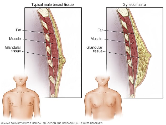
Enlarged Breasts In Men Gynecomastia Symptoms And Causes Mayo Clinic

Lymph glands in armpits and breast are important in cancer care and determination of the stage of breast cancer.

Swollen breast tissue. This swelling could be due to an inflammation which occurs as the bodys response to tissue injury. First 2 doctors I saw said I had a lump on my right breast above my nipple. Swollen clavicles refer to the appearance of a visible swelling or mass over the clavicular region.

This abnormal growth in breast tissueknown by the medical term gynecomastia often occurs in men over the age of 50 due to slowdown in the production of testosterone. The armpit has many lymph glands the lymph fluid from the breast tissue drains into these lymph nodes. Its normal for newborn babies boys and girls to have mild or even swollen enlarged breasts andor lumps under the nipple.

A breast lipoma is a lump formed by collection of fatty tissue within the breast. The COVID pandemic is resulting in delayed cancer diagnoses and will likely cause unnecessary deaths from breast cancer over the next several years Breast radiologists believe they will continue to see lymph node swelling as more and more patients become vaccinated so the Society of Breast Imaging has established recommendations to manage it in patients who have been vaccinated. The main cause of swollen breasts and tenderness is.

Premenstrual Syndrome PMS is a common cause of breast swelling. Breast swelling refers to the enlargement of one or both breasts typically accompanied by soreness or pain. Sometimes a swelling may actually be due to a mass like a tumors.

I was forwarded onto a surgeon and she examined me and told me it was more a mass of thickened skin tissue. As the estrogen levels increase in a womans body before the start of her periods the milk glands and breast tissues can swell due to water retention. Less commonly breast cancers can cause breast swelling particularly the type of breast cancer known as inflammatory breast cancer.

Swelling of the breast can occur as a response to infections or other causes of fluid buildup in the breast tissue. This is a normal process caused by hormonal changes 4 and can produce breast swelling and tenderness but probably wont cause breast pain. Mastitis is an infection of the breast tissue that results in breast pain swelling warmth and redness of the breast.

There are different types of breast cancer. I am 29 years old and after a trip to AE last night as my right breast was so swollen to my left. However as the periods end the size of breasts should return to normal.

Breast swelling can also be a symptom of breast cancer. In this case there is usually associated warmth pain or tenderness and redness of the breast. Mastitis most commonly affects women who are breast-feeding lactation mastitis although sometimes this condition can occur in women who arent breast-feeding.

The lumps are in the bodys lymph. Lymphatic Obstruction Lymphatic obstruction is the blockage in lymph flow within the breast and this may be due to a tumor infection injury or surgery and can cause a unilateral breast swelling. They are almost always benign and.

Some women are developing swollen lumps in their breasts as a result of the Covid-19 vaccine according to US doctors. A sensation of soreness typically occurs during menstrual periods pregnancy breastfeeding and menopause. Definition of Breast Swelling Breast swelling is an enlargement of the breast or both breasts compared to the regular breast size and may be accompanied by other symptoms like tenderness soreness pain lump s changes of the areola or nipple and any secretion from the nipple.

Inflammatory breast cancer can cause your breasts to.

Thursday, February 7, 2019

Breast Tissue Ultrasound

Breast tissues are either echogenic white or hypoechoic black on ultrasound. Breast ultrasound is an important modality in breast imaging.

Breast Ultrasound Radiology Reference Article Radiopaedia Org

Due to the dense fibroglandular tissue the tumor is not well seen.

Breast tissue ultrasound. Breast cancer grading and specific differentiation must involve a series of investigations and not be based on ultrasound alone. Breast tissue is composed of milk glands milk ducts and supportive tissue dense breast tissue and fatty tissue nondense breast tissue. 1 in the retroareolar region of both breast consisting of central echogenic tissue surrounded by numerous peripheral cysts of variable size.

The Breast Ultrasound Examination. Accessory breast tissue is a relatively common congenital condition in which abnormal accessory breast tissue is seen in addition to the presence of normal breast tissue. Arm above the head - helps thin and spread breast tissue for improved penetration Lying supine to evaluate medial tissue Lying supine oblique for lateral breast and axilla evaluation Wedges can be used to provide additional support.

No debris septations or fluid-fluid levels were seen. This is because it may miss some early signs of cancer. Ultrasound showed shrinkage of the tumor to a 18 mm mass which was categorized as BI-RADS 6.

Since the glandular tissue is more firm than the fatty tissue this feels like a mass on palpation. Breast ultrasound is not usually done to screen for breast cancer. The ultrasound uses high.

Indicates that the cancer is still contained entirely with the tissue of origin and not penetrated tissue boundaries a histological diagnosis. Hyperemia can be evidenced by Doppler ultrasound which reveals an increased number of arterial and especially venous structures. The ultrasound shows a collection of glandular tissue within a fatty breast.

A breast ultrasound is a scan that uses penetrating sound waves that do not affect or damage the tissue and cannot be heard by humans. What Is a Breast Ultrasound. Its a normal and common finding.

This leads to increased confidence in the tumor extent and location heading into treatment. Automated breast ultrasound can also provide vital details to prepare for surgery or other treatment and monitoring. The sound waves bounce off surfaces in your body and the.

The skin is an echogenic line immediately under the transducer in the near field. Ultrasound examination of the neonatal breast showed ovoid-shaped masses Fig. The breast tissue deflects these waves causing echoes which a computer uses to paint a picture of whats going on inside the breast tissue no radiation is involved.

Doctors often request the. Positioning is key to immobilize breast tissue and aid with scanning Good positioning includes. A mammogram was performed with a marker on the palpable mass and also showed a focal collection of normal glandular tissue.

A breast ultrasound is an imaging technique commonly used to screen for tumors and other breast abnormalities. For optimal high-resolution scanning of the breast with high frequency probes a basic principle is to minimize the thickness of tissue. In assessing for malignancy it is important to remember that one must use the most suspicious feature of three modalities pathology ultrasound and mammography to guide management.

When viewed on a mammogram women with dense breasts have more dense tissue than fatty tissue. After chemotherapy the tumor is not visible on the mammogram. Ultrasound demonstrated a 37 mm mass with indistinct and angular margins and shadowing.

A breast ultrasound is a painless procedure that uses sound waves to make images of the inside of your breast. There was no increased vascularity and the overlying skin was normal. 3D imaging helps clinicians more completely visualize tumors and surrounding tissue and provides more precise location information.

It is normally about 2 to 3 mm thick and has a hypoechoic layer of dark subcutaneous fat immediately beneath it Box 5-2. It is the usual initial breast imaging modality in those under 30 years of age in many countries ref. This normal variant can present as a mass anywhere along the course of the embryologic mammary.

Breast hyperemia defined as increased vascularization of breast tissue is also associated with inflammation. A breast ultrasound uses high frequency sound waves to create a black-and-white image of breast tissues and structures. Dense breast tissue refers to the appearance of breast tissue on a mammogram.

A breast ultrasound is most often done to find out if a problem found by a mammogram or physical exam of the breast may be a cyst filled with fluid or a solid tumor.

4 Month Appointment

The 4-month visit is a time to show off your beautiful baby. Then hell beam brightly and charm everyone around him. Pregancy Belly Month ...