Showing posts with label aortic. Show all posts

Thursday, February 4, 2021

Hypoplastic Aortic Arch

1 1988 66 consecutive neonates with coarctation and severe hypoplasia of the transverse arch underwent coarctation repair by resection of the coarctation and reconstruction of the aortic arch. Transverse arch hypoplasia is an integral albeit anatomically independent part of neonatal coarctation of the aorta.

Aortic Arch Anomalies Diagnosis Treatment Ssm Health

Patients with an interrupted or hypoplastic aortic arch usually present as neonates when the ductus arteriosus closes and flow to the descending aorta ceases or.

Hypoplastic aortic arch. In addition to narrowing and hypoplasia of the aortic isthmus with a posterior shelf segments of the transverse arch are also hypoplastic with z-scores of 3 or lower. Unlike coarctation a larger portion of the aorta is blocked and thus the condition is more serious and severe and requires a longer and more difficult operation to correct. Hypoplasia of aorta.

1 Extended end to end anastomosis has been advocated to overcome arch hypoplasia. Neonatal patients with hypoplasia of the aortic arch constitute a heterogeneous group with a wide spectrum of severity. This is the American ICD-10-CM version of Q2542 - other international versions of ICD-10 Q2542 may differ.

It means not coded here. Aortic arch anomalies are a type of congenital heart condition which means it is a disease or abnormality that is present from birth. Neonatal patients with hypoplasia of the aortic arch constitute a heterogeneous group with a wide spectrum of severity.

It can manifest as isolated stenosis or long-tubular hypoplasia of the transverse aortic arch TAA. The optimal treatment of neonates and infants with CoA and hypoplastic aortic arch HAA is controversial. 35 Limited information however is available on how rapidly the hypoplastic.

2 Numerous studies demonstrated the growth potential of the aortic arch following repair with confined extensity. A hypoplastic aortic arch may be associated with a ventricular septal defect and other congenital heart lesions. Aortic arch advancement for aortic coarctation and hypoplastic aortic arch in neonates and infants.

Q2542 is a billablespecific ICD-10-CM code that can be used to indicate a diagnosis for reimbursement purposes. The coarctation was isolated in 23 associated with a. Recurrent aortic arch obstruction and long-term hypertension remain significant problems 2 3 4.

63 of the newborn infants were less than 2 weeks of age. This anomaly results from regression of the right arch between the right common carotid and right subclavian arteries including the right ductus arteriosus Fig 6. Finding hope at CHOP.

The distal right dorsal aorta rather than the right fourth arch becomes the proximal right subclavian artery forming its retroesophageal portion. A type 1 excludes note indicates that the code excluded should never be used at the same time as Q254A type 1 excludes note is for used for when two conditions cannot occur together such as a congenital form versus an acquired form of the same condition. 1 1983 to Jan.

The milder end of the spectrum comprises patients with aortic coarctation and isthmus hypoplasia. The doctor told Natalie and her husband Joe that even if their baby survived to term he would probably die soon after birth. A type 1 excludes note is a pure excludes.

There are many different variations of aortic arch anomalies. In HLHS the hearts left side including the aorta aortic valve left ventricle and mitral valve is underdeveloped. Mean age at operation was 14 - 8 days ranging from 2 to 30 days.

The aorta arises from the left ventricle and carries oxygenated blood from the heart to the body. At the other end of the spectrum are patients with severe transverse arch hypoplasia or hypoplastic left heart syndrome. At her 20-week ultrasound during her third pregnancy Natalie found out that her unborn child had a condition called coarctation of the aorta with aortic arch hypoplasia.

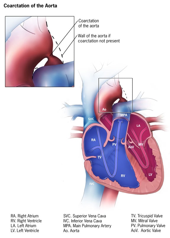
Coarctation of the aorta CoA is defined as congenital narrowing of the aortic isthmus near the ductus arteriosus or arterial ligament. Coarctation of the aorta can occur in combination with hypoplasia of the aortic arch. Traditionally diagnosis is made at cardiac catheterisation.

The 2021 edition of ICD-10-CM Q2542 became effective on October 1 2020. Coarctation of the aorta with concomitant hypoplastic aortic arch is a rare congenital anomaly affecting the cardiovascular system. Hypoplastic left heart syndrome HLHS is a heart condition present from birth congenital heart defect.

Cardiac catheterisation is an invasive procedure and therefore is fraught with complications especially when performed in ill infants. A condition similar to coarctation of the aorta a hypoplastic aortic arch is said to be present when there is a blockage in a certain location in the aorta. The milder end of the spectrum comprises patients with aortic coarctation and isthmus hypoplasia.

Some studies have shown variable degrees of growth of the proximal HAA after CoA repair through a left thoracotomy 5 6. At the other end of the spectrum are patients with severe transverse arch hypoplasia or hypoplastic left heart syndrome. CoA a common congenital heart disease CHD in clinical practice accounts for 68 of CHD.

Mery CM Guzmán-Pruneda FA Carberry KE Watrin CH McChesney GR Chan JG Adachi I Heinle JS McKenzie ED Fraser CD Jr Ann Thorac Surg 2014 Aug982625-33.

Sunday, November 10, 2019

Aortic Valve Disease

Aortic valve disease is a type of heart valve disease that occurs when the valve between your aorta the largest blood vessel and the left ventricle the hearts main pumping chamber doesnt work as it should. Aortic valve disease is common and has significant impact on prognosis and quality of life.

Aortic Stenosis Cardiosmart American College Of Cardiology

This prevents the valve from opening or closing completely.

Aortic valve disease. The aortic valve lies between the left ventricle and that aorta the largest blood vessel in the body that carries blood to the body from the heart. Abnormal valves produce turbulent blood flow and can be infected by bacteria. Aortic valve disease can be caused by aortic stenosis aortic regurgitation or a combination of both.

1 What does aortic valve disease mean. It has been shown to be an effective therapy in all age groups including the very elderly age 90 years. In this educational review we cover the pathophysiology presentation and assessment of aortic stenosis AS and aortic regurgitation AR including the role of imaging modalities beyond echocardiography.

36 Concept of the Heart Team and heart valve centres 37 Management of associated conditions 4. Aortic stenosis is a narrowing of the aortic valve opening. All patients with aortic valve disease should follow measures prescribed by their physician to prevent infection of the abnormal valve.

The valve can also become damaged by rheumatic fever infection or. Medical therapy 44 Serial testing 45 Special patient populations 5. In some cases the aortic valve is abnormal from birth.

Aortic valve disease may be a condition present at birth congenital heart disease or it may result from other causes. AORTIC DISEASEASAR BY DR NIKUNJ CTS RESIDENT STAR HOSPITAL CoordinatorDR PSATYENDRANATH PATHURI 21719 2. Aortic valve diseases AVD are often associated with a higher maternal and foetal risk.

The aortic valve may be abnormal from birth congenital or it could become diseased with age acquired. Leggett M1 Otto CM. There have been several significant advances in our understanding of aortic valve disease over the past year.

Many people have heart valve defects or disease but dont have symptoms. Aortic valve disease most often refers to obstruction of the aortic valve aortic stenosis or an aortic valve leak known as aortic regurgitation or aortic insufficiency. Acquired heart valve disease usually involves aortic or mitral valves.

What is aortic valve stenosis. Although the valves are normal at first problems develop over time. The physiological haemodynamic changes during pregnancy may not be well tolerated in patients with significant AVD.

Aortic valve disease 1. Aortic stenosis is one of the most common and serious valve disease problems. The most common congenital aortic valve abnormality called a bicuspid aortic valve occurs when the valve has only two leaflets bicuspid instead of three tricuspid.

Both congenital and acquired heart valve disease can cause stenosis or backflow. Aortic valve replacement is the most commonly performed valve operation. For some people the condition mostly stays.

That is why patients with AVD need pre-conception counselling to risk stratify their condition to ensure a safe and eventless pregnancy. Recent studies suggest that degenerative valvular aortic stenosis is an active disease process. Aortic Regurgitation 41 Evaluation 42 Indications for intervention 43.

ANATOMY The aortic valve is the last of four cardiac valves through which the blood is pumped before it goes to the rest of the body. Aortic stenosis restricts the blood flow from the left ventricle to the aorta and may also affect the pressure in the left atrium. The vast majority of aortic valve disease is an.

1Department of Medicine University of Washington Seattle 98195 USA. The most common etiologies for aortic stenosis are calcific degeneration rheumatic disease and. Aortic valve disease Aortic valve disease is a condition in which the valve between the main pumping chamber of your heart left ventricle and the main artery to your body aorta doesnt work properly.

Aortic Stenosis 51 Evaluation 52 Indications for intervention.

Monday, September 16, 2019

Heart Aortic Aneurysm

Incredibly the aorta pumps around 200000000 liters of blood around the body in a lifetime. If the bulging stretches the vessel wall too far the vessel may burst.

Aortic Aneurysm Uab Medicine

An aneurysm is a balloon-like bulge in the wall of a weakened blood vessel.

Heart aortic aneurysm. Aortic aneurysms can dissect or rupture. An aortic aneurysm is a bulge in the wall of the bodys largest artery the aorta. The aorta is the large blood vessel that the heart pumps into viathe aortic valve every time it beats.

Aortic disease or an injury may also cause an aneurysm. When the aortas walls become weakened or damaged the aortas ability to withstand this force decreases and the walls may bulge. An aneurysm occurs when part of an artery wall weakens allowing it to abnormally balloon out or widen.

They can also be hereditary. An aortic aneurysm is a balloon-like bulge in the aorta the large artery that carries blood from the heart through the chest and torso. An aortic aneurysm is a bulging or ballooning area in the wall of the aorta the bodys largest blood vessel which carries blood from the heart to the rest of the body.

The aortas walls are naturally elastic allowing them to absorb the pumping force of the heart. Read about the causes symptoms and the treatment of an aortic aneurysm. An aortic aneurysm is a localized dilation of the aorta the main artery in the body.

An aortic aneurysm occu. Also known as abdominal aortic aneurysm AAA thoracic aortic aneurysm TAA An aneurysm is a balloon-like bulge in an artery. Some people are born with them.

The aorta is the main pipe which takes oxygen-rich blood from your heart to the rest of your body. However other serious complications are also possible. The aorta is the artery that carries blood from the heart to the rest of the body.

If it becomes significantly large it can lead to catastrophic complications. An aortic aneurysm is an abnormal bulge that occurs in the wall of the major blood vessel aorta that carries blood from your heart to your body. Aortic aneurysms are aneurysms that occur in the aorta the main artery carrying oxygen-rich blood to.

It is the largest artery in your body. Aortic aneurysms can occur anywhere in your aorta and may be tube-shaped fusiform or round saccular. Other factors include High blood pressure High blood cholesterol Atherosclerosis hardened arteries Some inherited connective tissue disorders such as Marfan.

A new study investigates a genetic culprit behind abdominal aortic aneurysm a serious condition that puts people at risk of their aorta rupturing -- a potentially deadly event. The chief reason it is considered a problem is that sometimes aneurysms can rupture leading to catastrophic internal bleeding. The causes of aneurysms are sometimes unknown.

Diseases and unhealthy behaviors that damage your heart and blood vessels also increase your risk for aortic aneurysm. An aneurysm in the aorta is called an aortic aneurysm. If an aortic aneurysm a bulge in the wall of your bodys main arteryis larger than 2 inches or 50 to 55 centimeters in diameter is growing fast or is causing serious symptoms such as pain or trouble breathing it is advisable to consider the possibility of surgical repair.

The aneurysm forms in the wall of the artery. Arteries are blood vessels that carry blood from your heart to your organs. An aortic aneurysm is a balloon-like bulge that affects the aorta the main artery that carries oxygen-rich blood directly from the heart to smaller blood vessels in your body.

Smoking is the most important behavior related to aortic aneurysm. What are the risk factors for aortic aneurysm. The aorta is the largest artery in the body and transports oxygen-rich blood from the hearts left ventricle to the rest of the body.

Aortic aneurysmdescribes an aorta that is enlarged in size. The force of blood pumping can split the layers of the artery wall allowing blood to leak in between them.

Friday, August 3, 2018

Aortic Rupture Symptoms

The condition is caused by trauma to the aorta. The greatest danger is the rupture of the aortic aneurysm.

Aortic Dissection Causes Symptoms Complications And Treatments

It can be tormented by eructations urination disorders constipation.

Aortic rupture symptoms. Symptoms of rupture usually begin with sudden severe pain in the chest or back followed rapidly by palpitations severe weakness and lightheadedness shock and loss of consciousness. Aortic aneurysm is an extremely dangerous disease and its rupture is deadly therefore it is important to detect pathology in time and if possible carry out planned treatment. Cardiovascular collapse is usually so rapid that even if an attempt can be made to surgically repair the rupture mortality is excessively high.

Severe chest abdominal or back pain. The most common symptoms of an aortic aneurysm rupture are not necessarily similar to those of an un-ruptured aortic aneurysm although the pain in the area of an aneurysm can occur. If you suspect that you or someone else has had a ruptured aneurysm call 999 immediately and ask for an ambulance.

Participating in certain activities or lifestyle choices could increase the risk of experiencing traumatic aortic transection. The prognosis remains serious mortality at breaks reaches 90 while planned treatment gives a good result and the probability of death does not exceed 5. People at risk include those with a history of aortic aneurysm or blockages of other blood vessels and patients with high blood pressure.

When present symptoms of an aortic rupture may include. Abdominal aortic aneurysm rupture is most commonly presented in a classic triad of symptoms seen in about one half of patients. Read more about the symptoms of an AAA.

The symptoms of aortic dissection include. Signs of external chest injury. Signs may include asymmetric pulses or blood pressure decreased blood flow to the lower extremities and precordial systolic murmur.

This is an unpleasant but not the most dangerous condition. Severe chest pain Severe back pain Severe abdominal pain Signs of external chest injury Causes and Risk Factors The condition is caused by trauma to the aorta. The aorta can rupture completely or incompletely after blunt or penetrating chest trauma.

Signs Symptoms of Ruptured Aorta Patient experiences tearing pain in the abdomen back flank or groin. Causes of an AAA. Abdominal or back pain which is sudden sharp and severe.

Lightheadedness dizziness and blurred vision. Fluoroquinolones may cause aortic rupture in some CIDRAP. When present symptoms of an aortic rupture may include.

A sudden onset of severe pain across the chest often felt in the back or between the shoulder blades pain in the jaw face abdomen back or lower extremities feeling cold clammy and sweaty. What are the symptoms of an AAA rupture. With an abdominal aneurysm a person feels pain in the epigastric region as well as a sensation of a strong swelling of the abdomen nausea.

Causes and Risk Factors. Hypotension low blood pressure. The most common symptom of a ruptured aortic aneurysm is sudden and severe pain in the abdomen.

4 Month Appointment

The 4-month visit is a time to show off your beautiful baby. Then hell beam brightly and charm everyone around him. Pregancy Belly Month ...