Breast tissues are either echogenic white or hypoechoic black on ultrasound. Breast ultrasound is an important modality in breast imaging.
 Breast Ultrasound Radiology Reference Article Radiopaedia Org
Due to the dense fibroglandular tissue the tumor is not well seen.
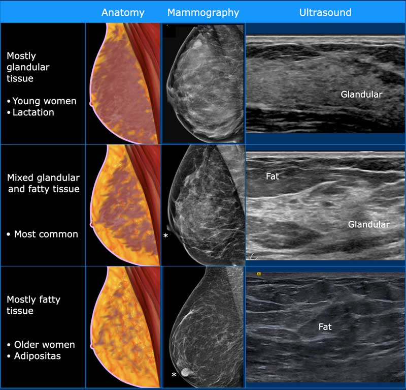
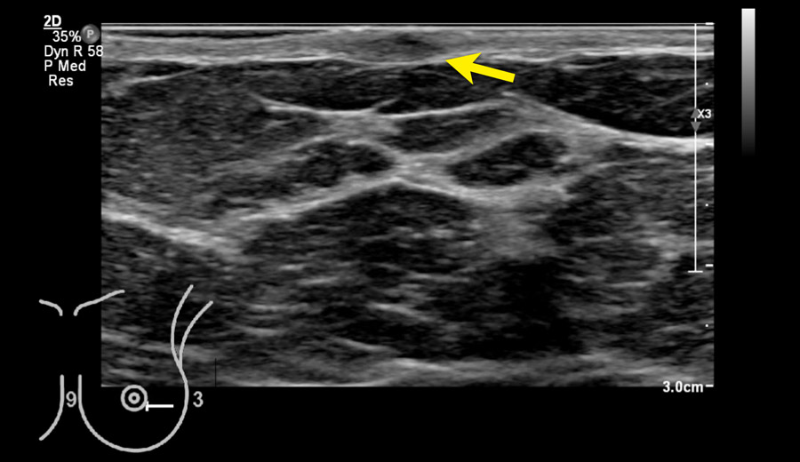
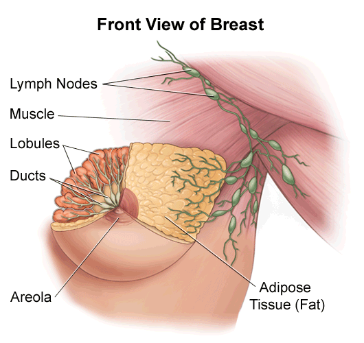
Breast tissue ultrasound. Breast cancer grading and specific differentiation must involve a series of investigations and not be based on ultrasound alone. Breast tissue is composed of milk glands milk ducts and supportive tissue dense breast tissue and fatty tissue nondense breast tissue. 1 in the retroareolar region of both breast consisting of central echogenic tissue surrounded by numerous peripheral cysts of variable size.
The Breast Ultrasound Examination. Accessory breast tissue is a relatively common congenital condition in which abnormal accessory breast tissue is seen in addition to the presence of normal breast tissue. Arm above the head - helps thin and spread breast tissue for improved penetration Lying supine to evaluate medial tissue Lying supine oblique for lateral breast and axilla evaluation Wedges can be used to provide additional support.
No debris septations or fluid-fluid levels were seen. This is because it may miss some early signs of cancer. Ultrasound showed shrinkage of the tumor to a 18 mm mass which was categorized as BI-RADS 6.
Since the glandular tissue is more firm than the fatty tissue this feels like a mass on palpation. Breast ultrasound is not usually done to screen for breast cancer. The ultrasound uses high.
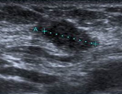
Indicates that the cancer is still contained entirely with the tissue of origin and not penetrated tissue boundaries a histological diagnosis. Hyperemia can be evidenced by Doppler ultrasound which reveals an increased number of arterial and especially venous structures. The ultrasound shows a collection of glandular tissue within a fatty breast.
A breast ultrasound is a scan that uses penetrating sound waves that do not affect or damage the tissue and cannot be heard by humans. What Is a Breast Ultrasound. Its a normal and common finding.
This leads to increased confidence in the tumor extent and location heading into treatment. Automated breast ultrasound can also provide vital details to prepare for surgery or other treatment and monitoring. The sound waves bounce off surfaces in your body and the.
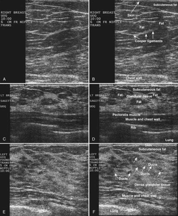
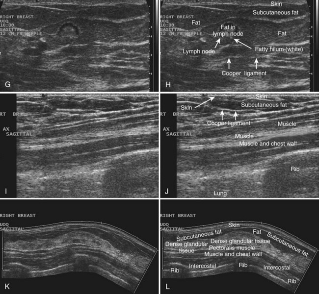
The skin is an echogenic line immediately under the transducer in the near field. Ultrasound examination of the neonatal breast showed ovoid-shaped masses Fig. The breast tissue deflects these waves causing echoes which a computer uses to paint a picture of whats going on inside the breast tissue no radiation is involved.
Doctors often request the. Positioning is key to immobilize breast tissue and aid with scanning Good positioning includes. A mammogram was performed with a marker on the palpable mass and also showed a focal collection of normal glandular tissue.
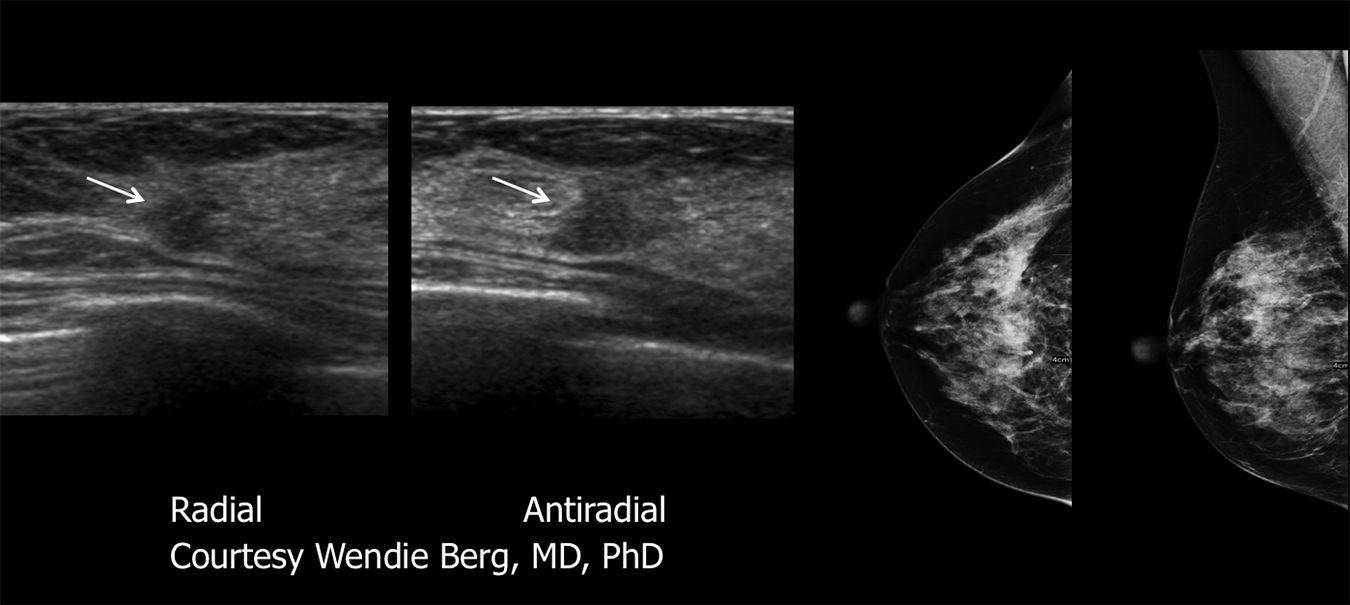
A breast ultrasound is an imaging technique commonly used to screen for tumors and other breast abnormalities. For optimal high-resolution scanning of the breast with high frequency probes a basic principle is to minimize the thickness of tissue. In assessing for malignancy it is important to remember that one must use the most suspicious feature of three modalities pathology ultrasound and mammography to guide management.
When viewed on a mammogram women with dense breasts have more dense tissue than fatty tissue. After chemotherapy the tumor is not visible on the mammogram. Ultrasound demonstrated a 37 mm mass with indistinct and angular margins and shadowing.
A breast ultrasound is a painless procedure that uses sound waves to make images of the inside of your breast. There was no increased vascularity and the overlying skin was normal. 3D imaging helps clinicians more completely visualize tumors and surrounding tissue and provides more precise location information.
It is normally about 2 to 3 mm thick and has a hypoechoic layer of dark subcutaneous fat immediately beneath it Box 5-2. It is the usual initial breast imaging modality in those under 30 years of age in many countries ref. This normal variant can present as a mass anywhere along the course of the embryologic mammary.
Breast hyperemia defined as increased vascularization of breast tissue is also associated with inflammation. A breast ultrasound uses high frequency sound waves to create a black-and-white image of breast tissues and structures. Dense breast tissue refers to the appearance of breast tissue on a mammogram.
A breast ultrasound is most often done to find out if a problem found by a mammogram or physical exam of the breast may be a cyst filled with fluid or a solid tumor.
 Normal Breast Ultrasound How To
 Breast Ultrasound Clinical Gate
 Breast Ultrasound Past Present And Future Intechopen
 Weinstein Imaging Associates Dbtust
 The Radiology Assistant Ultrasound Of The Breast
 The Radiology Assistant Ultrasound Of The Breast
 Breast Radiology Key
 Ultrasound Of Normal Right Sided Breast Tissue Left Top Left Side Download Scientific Diagram
 The Good The Bad And The Ugly About Breast Ultrasound
 Breast Ultrasound Clinical Gate
 Normal Breast Ultrasound How To
 Breast Ultrasound Sonogram Densebreast Info Inc
 Ultrasound Image Of The Lactating Breast The Skin Is Displayed Download Scientific Diagram


No comments:
Post a Comment
Note: Only a member of this blog may post a comment.