Showing posts with label brain. Show all posts

Monday, September 28, 2020

Treatment For Ischemic Changes In The Brain

Medications to manage high blood pressure reduce cholesterol andor thin the blood may be prescribed to treat cerebral ischemia as well as having the individual quit smoking participate in an exercise routine change poor eating habits and manage. These are signs of microvascular ischemic disease in the brain tissue.

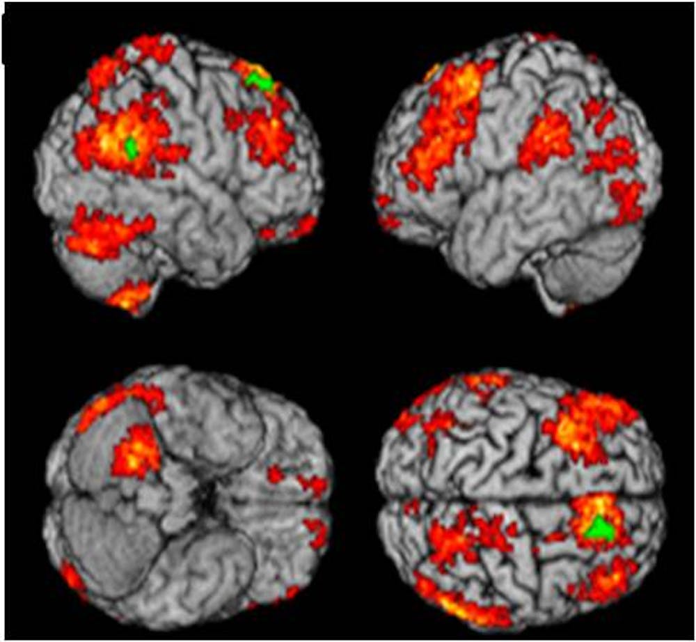
Update On Cerebral Small Vessel Disease A Dynamic Whole Brain Disease Stroke And Vascular Neurology

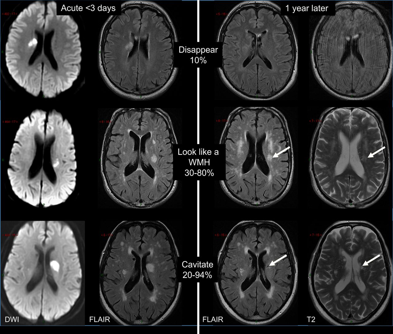
If the patient is given and infusion of gadolinium the spots will appear even brighter on the MRI film.

Treatment for ischemic changes in the brain. It means that the tiny blood vessels in the brain are blocked. 27 years experience Neurosurgery. Treatment generally involves managing the risk factors that contribute to small blood vessel damage in the brain.

MRI of the Brain with Ischemic Changes. Treating microvascular ischemic disease When a person experiences a mild case treatment focuses on lifestyle adjustments that should improve overall health. Endovascular treatment such as balloon angioplasty and implanting stents may be performed which open the narrowed or blocked blood vessels.

The first target in the treatment is to regulate the breathing by normalizing the heart rate and blood pressure. Have it checked in both arms. What does microvascular ischemic changes mean.

If left untreated a part of the brain tissue can die off. Dissolving a blood clot is one treatment for an ischemic stroke. Learn what causes it what the symptoms are and how you can prevent it.

Cerebral or brain ischemia is a condition that occurs when there isnt enough blood flow to the brain. It is incidentally picked up in a brain scan such as MRI magnetic resonance imaging. Which treatment strategy your doctor recommends will depend on.

Treatment of ischemia Treatment depends on personal circumstances such as age and medical history. These individuals may have to change. Ischemia is a serious problem where some part of your body like your heart or brain isnt getting enough blood.

Depending on the severity of your condition you may be treated with medications surgery or both. The treatment a health care provider will choose for ischemic stroke will depend on the location and size of the clot. More likely it is your high blood pressure that is causing the ischemic damage to your brain and your headache.

In this MRI of the brain there are multiple white spots that appear very brightly in the brain tissue. Chronic Microvascular Ischemic Disease MRI Sample. This MRI was done for a 62-year old woman who suffered from migraine headaches.

As we get older arteries harden and we see small vessel changes in the brain due to aging. Smaller blood clots may be removed using a medication which dissolves the clot itself or thins the blood. Treatment approaches may include lifestyle changes and medications to reduce the risk of stroke physical disability and cognitive decline.

Doctors at Columbia Neurosurgery in New York will discover the symptoms and causes In order to successfully treat cerebral ischemia. Medications used to treat various types of ischemia include anti-platelet drugs and anticoagulants. Doctors choice of management may depend on the patients symptoms and risk factors best suiting a patient of small vessel ischemic disease.

Treatment options include lifestyle changes and medications to help reduce the risk of stroke cognitive decline and physical disabilities. A doctor may recommend one or. An MRI was done four years prior with some of the changes see above.

If necessary your doctor will then try to reduce pressure in the brain with medication. If that shows signs of a problem an MRI and or a CT scan will be ordered to see the changes inside the blood vessels in the brain. They simply mean the brain doesnt work as well as it did.

They are usually filled with clots but in some instances appear to be ruptured. Whenever possible a neurosurgeon will recommend conservative treatment which would address the narrowed arteries with medicine and lifestyle changes. 57 years experience Endocrinology.

The main treatment for ischemic stroke is intravenous tissue plasminogen activator tPA which breaks up.

Monday, August 3, 2020

Human Brain Development

These include a cerebral cortex containing progenitor populations that organize and produce mature cortical neuron subtypes. Epigenetics and Genetics of Development.

Building A Human Brain In Miniature By Just A Brain Thing Medium

The developing brain goes through many stages.

Human brain development. If you are constantly under duress andor have severe anxiety it is best to take steps to minimize it. Emotional well-being and social competence provide a strong foundation for emerging cognitive abilities and together they are the bricks and mortar that comprise the foundation of human development. At birth a persons brain will have almost all the neuronsthat it.

Your babys brain will triple in size during this time growing from a little over 3 ounces to almost 11 ounces at birth. At times during brain development 250000 neuronsare added every minute. The Science of Early Childhood Development This brief explains how the science of early brain development can inform investments in early childhood and helps to illustrate why child developmentparticularly from birth to five yearsis a foundation for a.

For men they become fully mature at age 18. At birth the average babys brain is about a quarter of the size of the average adult brain. In human development a recent study shows that slower microstructural development between 27 and 46 weeks post conception 2948 weeks gestational age in premature infants was related to lower levels of neurodevelopmental functioning at the age of 2 years 31.

Brain development is shaped by the integration. Brain development in the third trimester is marked by the rapid development of neurons in the brain and explosive growth. The brain is the command center of the human body.

It consists primarily of the cerebrum 2 and the structures hidden beneath it see The Inner Brain. Cortical grey matter development peaks at 12 years of age in the frontal and parietal cortices and 17 years in the temporal lobes with the superior temporal cortex being last to mature for women and they have reached full maturity at age 16-17. The human brain begins forming very early in prenatal life just three weeks after conception but in many ways brain development is a lifelong project.

The forebrain is the largest and most highly developed part of the human brain. The development of the nervous system in humans or neural development or neurodevelopment involves the studies of embryology developmental biology and neuroscience to describe the cellular and molecular mechanisms by which the complex nervous system forms in humans develops during prenatal development and continues to develop postnatally. The cerebrum will begin to develop grooves and ridges and separate into the left brain and right brain.

In a 1974 survey of data on human brain and head growth statistically significant peaks in growth rates of human brains were found around ages 11 years and 15 years. When people see pictures of the brain it is usually the cerebrum that they notice. In the embryos of vertebrates the predecessor to the brain and spinal cord is the neural tube.

It is recommended to avoid exposure to illicit. Proper nutrition starting in pregnancy Exposure to toxins or infections The childs experiences with other. It keeps growing to about 80 of adult size by age 3 and 90 nearly full grown by age 5.

By age six the brain is 95 its adult weight and peak of energy consumption. The brain is a highly interrelated organ and its multiple functions operate in a richly coordinated fashion. Some landmarks of neural development.

Drinking excessive amounts of alcohol to the point of abuse may have an impact on the developing brain. Amazing rate during development. Caregivers need to provide nurturing environment and daily.

Here we have developed a human pluripotent stem cell-derived three-dimensional organoid culture system termed cerebral organoids that develop various discrete although interdependent brain regions. Thanks to the latest technologies though we can now trace the brains evolution in unprecedented detail from a time before the very first nerve cells right up to the age of cave art and cubism. How well a brain develops depends on many factors in addition to genes such as.

As the fetus develops the grooves and folds in the neural tube deepen giving rise to different layers of the brain. Brain Development Brain Development. That is because the same events that shape the brain during development are also responsible for storing informationnew skills and.

5 Stages of Human Brain Development Development of voluntary movement reasoning perception frontal lobes active in development of emotions attachments. Hemispheric Asymmetries Over the. Incredibly it doubles in size in the first year.

Brain development has a protracted time course in humans beginning in the third week post conception.

Saturday, May 9, 2020

What Does A Depressed Brain Look Like

People who have never known what that feels like try to be helpful saying things like this too shall pass or get over it or dont worry be happy. If only it were that easy.

Brain Areas Related To Cognitive Effects Of Depression Brain Image Download Scientific Diagram

When youre depressed you remember think suffer and reason in search of solutions and new alternatives.

What does a depressed brain look like. While we know the neurochemicals involved and many contributing factors the truth is we really dont know the. The amygdala is a brain structure that is typically associated with regulating emotions. The amygdala is activated when a person recalls emotionally charged memories such as a frightening situation.

Reduced functionality of the hippocampus. Last medically reviewed on June 25 2017. The depressive stage of BMD may look a lot like major depression.

We found that there is a widespread network of brain regions which includes specific density variations in both grey and white matter in patients with depression. Your clinician should consider an evaluation for BMD if any of the following exist alongside the manic symptoms described above. Depression is being pulled into darkness and forgetting that light ever existed.

The effects of depression on the brain also can result in structural and connective changes. A study published in Molecular Psychiatry today has proved once and for all that recurrent depression shrinks the hippocampus - an area of the brain responsible for forming new memories -. Even well-trained mental-health professionals struggle to separate them especially in the early years of depressive symptoms.

Its also harder to sleep because youre thinking about so many different things. This can result in memory impairment. What Does the Depressed Brain Look Like.

What does a depressed brain look like. A depressed brain makes the hippocampus and prefrontal cortex smaller enlarged amygdala and thicker gray matter. Your hippocampus is therefore active which is the front part of the brain.

The evidence affirms that depression truly is a biologically based disorder of the brain and left unchecked it may run a degenerative course that damages brain tissue in ways similar to other. In the study published today Nov. The hippocampus is the part of the brain that regulates memory and emotion.

That kind of fight takes major strength. People with depression are often found to have a more active amygdala than in a normal brain. If visible differences do exist they are usually seen in the frontal cortex or hippocampus.

Its not simply a weakness that somebody should get over or even something we have a say in the narrator says. Areas of the brain affected by depression. Can depression be seen in the brain.

On rare occasions someone could have a structural issue that results in depression like a tumor or brain cyst. F undamentally a depressed brain looks just like any other brain. They looked for differences in the gray matter of the brain focusing specifically on the thickness of the cerebral cortex a thick outer layer of tissue that is densely packed with neurons and.

Its easy to understand. Depression is not darkness without hope for light. In fact theres no brain scan MRI EEG or any other medical test that can definitively diagnose depression and its not understood in detail like other brain disorders.

The size of this structure has been found to decrease in chronically depressed people. In particular the amygdala in depressed people is more active than in people without depression when exposed to a negative stimulus such as a sad face. In most cases a static brain scan of a depressed person looks the same as the brain scan of someone without depression.

If were being totally honest the people who fight depression and its symptoms sounds like the opposite of weak. The amygdala is part of the limbic system a group of structures deep in the brain thats associated with emotions such as anger pleasure sorrow fear and sexual arousal. 8 in the journal Cell a group of.

Feelings of sadness or anxiety might be linked to increased chitchat between two areas of the brain a new study suggests.

Sunday, September 1, 2019

Epilepsy Brain Scan

Magnetic Resonance Imaging MRI scan. Now scientists have developed a brain scanning technique that can find the source of a patients fits meaning they can potentially be cured surgically.

Seizure Induced Brain Lesions A Wide Spectrum Of Variably Reversible Mri Abnormalities European Journal Of Radiology

The rest of the time the brain activity may be entirely normal.

Epilepsy brain scan. Brain imaging with MRI identifies structural cerebral pathology that may give rise to seizures. Ad Full Body Comprehensive CT Scan Based in Maryland. But for many people a brain scan does not show a cause for their seizures and even if no physical cause is seen the person may still have epilepsy.

Structural imaging reveals most cerebral lesions underlying focal epilepsy. Second we evaluated the whole-brain functional connectivity of the visual thalamic nuclei in the various populations of subjects under investigation. An appropriate angle must be given in the axial plane perpendicular to mid line of the brain.

The scan produces pictures of the brain which might show a physical cause for epilepsy such as a scar on the brain. Ad Full Body Comprehensive CT Scan Based in Maryland. In the process they can injure themselves and the condition can seriously affect a persons quality of life.

X-ray CT scanning has a role in assessing patients with seizures. PET scan may show abnormalities even if the brain MRI is normal. The greatest yield is from MRI at 3T using epilepsy protocols and reported by expert neuroradiologists who possess the full clinical data.

A brain scan can help spot problems in your brain that can sometimes cause epilepsy such as. Common diagnostic tests include electroencephalography EEG which measures electrical activity in the brain and brain scans such as magnetic resonance imaging MRI or computed tomography CT. In patients with epilepsy decreased brain function is seen in the region where seizures originate when the patient is not actually having a seizure.

Which is pretty important when trying to identify epilepsy or indeed many other brain issues. An EEG test gives information about the electrical activity that is happening in your brain at the time the test is carried out. In the United States an estimated 34 million people have epilepsy.

Forty-four patients with epilepsy and 16 healthy control subjects underwent an electroencephalography-correlated functional magnetic resonance imaging study during an eyes-closed condition. Brain imaging is fundamental to the evaluation of people with epilepsy to identify any underlying structural brain abnormality that may be the cause of the epilepsy and to identify a treatment need such as for a tumour vascular disease or infection. However these scans are incredible and play a key part in helping diagnose and treat epilepsy.

An unusual growth brain tumour damage to the brain such as damage caused by a. An MRI scan looks at the structure of the brain and may help to find the cause of. T1 tse coronal oblique 2mm epilepsy protocol Plan the coronal high resolution slices on the sagittal plane.

Angle the position block plane perpendicular to the long axis of the hippocampus. Check the positioning block in the other two planes. Many people with epilepsy only have unusual electrical activity in their brain when they are having a seizure.

Epilepsy is a brain condition that causes patients to fit and lose consciousness. Some of the scans can be noisy MRI fMRI the EEG needs your scalp to be scratched a bit sounds strange right and some need you to go into a little tunnel for a period of time all scans bar the EEG and MEG. Brain imaging has a crucial role in the presurgical assessment of patients with epilepsy.

On the other hand if the patient has a seizure during the test increased brain function is seen.

Wednesday, June 12, 2019

Water On The Brain In Adults

A stroke may be the cause of water on the brain. Hydrocephalus is a condition in which excess cerebrospinal fluid CSF builds up within the ventricles fluid-containing cavities of the brain and may increase pressure within the head.

Normal Pressure Hydrocephalus Nph

In the case of brain injury the traumatic injury or bleeding in the brain may increase the risk of developing hydrocephalus.

Water on the brain in adults. Although hydrocephalus was once known as water on the brain the water is actually cerebrospinal fluid CSF--a clear fluid that surrounds the brain and spinal cord. The body typically produces enough CSF each day and absorbs the same amount. Sheth Teaches Practical Deep Brain Stimulation Seminar Big News from US News World Report 2014 Best Hospital Survey Blair Ford MD Boy Has Life Saving Brain Surgery and.

Hydrocephalus that develops in adults and children acquired hydrocephalus is often caused by an illness or injury that affects the brain. Hydrocephalus that develops in older people normal pressure hydrocephalus may also be the result of an infection illness or injury but in many cases its not clear what causes the condition. If left untreated hydrocephalus can be fatal.

Normally cerebrospinal fluid flows through the ventricles and bathes the brain and spinal cord before being reabsorbed into the bloodstream. The excess fluid puts pressure on the brain which can damage it. Meyers Co-Chairs a New Study Beyond Theory.

Hydrocephalus also referred to as fluid or water on the brain is a condition where too much cerebrospinal fluid has built up in the skull. The build-up is often. For example lesions or tumors that appear in the brain and spinal cord may contribute to water on the brain in adults.

Hydrocephalus is a build-up of fluid in the brain. Hydrocephalus is a condition that occurs when fluid builds up in the skull and causes the brain to swell. Although hydrocephalus often is described as water on the brain the water is actually CSF a clear fluid surrounding the brain and spinal cord.

Winfree Looks at Ways to Answer the Question Best Doctors in NY now Available to More in NJ Best Endovascular Treatments. Cerebrospinal fluid CSF is the clear colorless fluid that protects and cushions the brain and spine. However many cases of hydrocephalus in adults occur without a history of these preceding illnesses.

Sometimes referred to as water on the brain NPH is a buildup of cerebrospinal fluid CSF in the brain of older adults that leads to a gradual deterioration of motor skills andor cognitive function. In contrast adults with hydrocephalus were usually born with normal brain anatomy but acquired a blockage due to a tumor injury bleed or infection. The CSF puts pressure on structures within the brain causing symptoms.

The excessive accumulation of CSF results in an abnormal widening of spaces in the brain called ventricles. When diagnosed correctly procedures to drain the excess CSF fluid can be performed and patients can often return to a normal life. Cerebral edema swelling that occurs in the brain is a severe and potentially fatal complication of stroke.

Hydrocephalus also called water in the brain is a condition where there is an abnormal build up of CSF cerebrospinal fluid in the cavities ventricles of the brain. New research shows for the first time that the glymphatic system -. Symptoms may start out mild and become more severe as more CSF accumulates.

Symptoms of hydrocephalus depend on the area of the brain affected and the severity of the hydrocephalus. The name means water on the brain Brain damage can occur as. The damage to the brain from hydrocephalus can cause a wide range of symptoms including.

In noncommunicating hydrocephalus the circulation of the fluid is blocked. Other factors that may lead to adult hydrocephalus are infections such as bacterial meningitis and mumps. The increase of spinal fluid takes up space in the.

When someone has the form of water on the brain known as communicating hydrocephalus the cerebrospinal fluid can circulate successfully between the ventricles of the brain but it cannot drain properly.

Sunday, January 6, 2019

Types Of Brain Diseases

Alzheimers disease is an age-related irreversible brain disorder that develops over a period of years. Dementia is another very dreadful brain disease that attacks the elderly people.

Types Of Brain Diseases With Their Description And Symptoms Download Scientific Diagram

The brain is the center of the nervous system and any disease in brain cripples our neuro-transmission.

Types of brain diseases. Vascular contributions to cognitive impairment and dementia Mixed dementia a combination of two or more types of dementia Other conditions that cause dementia-like symptoms can be halted or even reversed with treatment. Usually in encephalitis and meningitis infection is not confined to one area. Researchers have still not found the proper reason behind the formation of these tumors.

Risk factors are different for different types of brain disorders. Further brain tumors have been classified as grades 123 and 4. Some of the major categories of brain diseases are inflammation trauma tumours stroke seizures etc.

Some common types of brain disorders are listed such as Alzheimers Disease brain injuries seizure stroke autism bipolar and schizophrenia brain disorder. Brain diseases in the category of infections which can be classified into different forms. Types of brain tumors are masses of malignant cells that can grow in the brain or its envelopes.

Cerebro-Oculo-Facio-Skeletal Syndrome COFS National Institute of Neurological Disorders and Stroke Corticobasal Degeneration National Institute of Neurological Disorders and Stroke Gerstmann-Straussler-Scheinker Disease National Institute of Neurological Disorders and Stroke Kuru National Institute of Neurological Disorders and Stroke. Are generally divided into two categories. Brain disorders can affect anyone.

Headache is the most common type of brain disease which can take the form of migraine where the pain in brain is so excessive that the person feels very sick. While the malignant tumor is cancer forming benign tumors are non-cancerous. Some brain diseases such as Tay-Sachs disease Huntingtons disease muscular dystrophies and Wilson disease are genetic.

Aicardi syndrome only affects females and in very rare cases males with Klinefelter syndrome XXY. List of neurological conditions and disorders From Wikipedia the free encyclopedia This is a list of major and frequently observed neurological disorders eg Alzheimers disease symptoms eg back pain signs eg aphasia and syndromes eg Aicardi syndrome. However infection that affects mainly the meninges is usually called meningitis and infection that affects mainly the brain is usually called encephalitis.

Other types of progressive brain disease include. Disorders of all types are addressed in this detailed brain diseases list. Traumatic brain injury TBI.

Dementia Alzheimers and LBD. Traumatic brain injury is most common in children. Technically when both the brain and the meninges are infected the disorder is called meningoencephalitis.

Other types of defects of the brain such as microcephaly small brain. Some of the genetic brain disorders include leukodystrophies phenylketonuria Wilson disease and Tay-Sachs disease know their symptoms and treatment. There are many different types of brain diseases including Alzheimers Disease Dementias Brain Cancer Epilepsy Seizure Disorders and mental disorders.

Those suffering from brain neurological diseases can have symptoms ranging from seizures and physical affects to erratic behavior and mood swings. Alzheimers is the most common cause of dementia a group of disorders characterized. Parkinsons and Other Movement Disorders Stroke and Transient Ischemic Attack TIA.

Or porencephalic cysts a gap in the brain where there should be healthy brain tissue. Many brain disorders are genetic while a small number can be brought on by environmental factors. 1 Primary tumors of the brain which develop from brain cells and 2 brain metastases.

Let us have a look into some of the most common brain diseases along with their symptoms causes and their treatments. There are two types of brain tumors Malignant and Benign.

Saturday, December 29, 2018

Storming Brain Injury

Severe TBT is correlated with an exaggerated stress response due to plasma catecholamine levels known as sympathetic storming. Traumatic Brain Injury TBI is one of the leading causes of death.

Storming After Brain Injury Why Neurostorming Happens

After a severe brain injury your loved one may remain in a coma for some time.

Storming brain injury. A sympathetic storm is related to a set of distressful symptoms. The film will offer an intimate look into Ryans perpetual care regimen crafted by his innovative father Ken whos devoted his lifes work to. When the SNS is triggered your body releases adrenaline to prepare the body for fighting or fleeing.

This is a series of actions andor words given off by someone as their brain is re. Sympathetic storming after traumatic brain injury. A term commonly used by.

In fact comas are considered the first stage in traumatic brain injury recovery as they allow the brain to begin its healing process uninterrupted. Storming is a medical phenomenon that follows a brain injury. Over 85 Million Visitors.

It is caused by an injury to the upper portions of the brain stem or the hypothalamus responsible for the autonomic nervous system ANS - see site encyclopaedia. This is not always a negative sign. Fast heart rate fast breathing rate high blood pressure fever sweating increased muscle tone This syndrome frequently called storming can be very frightening for families and the.

Storming 1 After someone has suffered a moderate-to-severe brain injury they may develop a group of symptoms such as. It is also autonomic dysfunction syndrome. The SNS controls your bodys fight-or-flight response that activates in the presence of imminent danger.

After a Traumatic Brain Injury an individual will most likely Neurostorm. Following acute multiple trauma hypothalamic stimulation of the sympathetic nervous system and adrenal glands causes an increase in circulating corticoids and catecholamines or a stress response. Storming after brain injury refers to an excessive response of the sympathetic nervous system SNS.

The medical term for this is sympathetic storm. The onset of sympathetic storming should trigger the institution of scheduled enteric medications to provide continuous dampening of activity of the sympathetic nervous system. Ad Traumatic Brain Injury Lawyer Search Now.

Over 85 Million Visitors. Storming can occur within the first 24 hours after injury or up to weeks later1 The precise mechanism for the increase in activity of the sympa-thetic nervous system is unknown but the increased activity is thought to be a stage of recovery from severe traumatic brain injury2 Terms used to describe this phe-nomenon in published reports. In individuals with severe traumatic brain injury or a Glasgow Coma Scale score of 3-8 this response can be exaggerated and episodic.

Dysautonomia dys-auto-nomia is a syndrome that is also referred to among many other names as storming or autonomic dysfunction syndrome. A coma is the deepest state of unconsciousness. PSH also called neurostorming is often experienced by patients in the earlier phases of their recovery.

This phenomenon is also associated with brain tumors severe hydrocephalus and subarachnoid hemorrhage. In Storming we will follow the daily lives and challenges of Ken and Sue Diviney 10 years following a tragic attack which left their son Ryan comatose with a severe traumatic brain injury TBI. Ad Traumatic Brain Injury Lawyer Search Now.

Storming happens to be the mercifully succinct moniker given to Paroxysmal Sympathetic Hyperactivity PSH a nervous system disorder affecting 15 to 33 percent of individuals who have suffered a traumatic brain injury. Unfortunately because the brain itself is injured it difficult for the person to inhibit this sympathetic surge often called paroxysmal sympathetic hyperactivity PSH. Riding out the storm.

4 Month Appointment

The 4-month visit is a time to show off your beautiful baby. Then hell beam brightly and charm everyone around him. Pregancy Belly Month ...