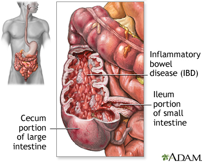
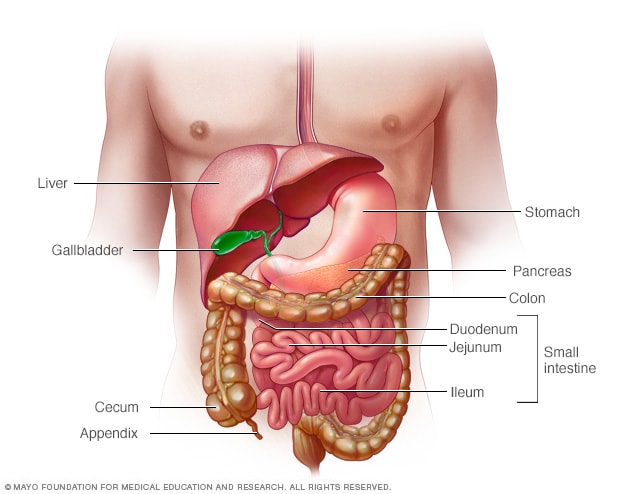
Anatomically the small bowel is divided into the duodenum the jejunum and the ileum. Small bowel obstruction is a partial or complete blockage of the small intestine which is a part of the digestive system.
 Crohn Disease Medlineplus Medical Encyclopedia
Celiac disease is an inflammatory small bowel disease triggered by gluten ingestion and typically resulting in diarrhea predominant symptoms but can be asymptomatic and detected by screening for antibodies against tissue transglutaminase.

Small bowel disease. Diverticula can occur in any portion of the gastrointestinal GI tract. A diverse array of disorders can be diagnosed with small bowel biopsy. Here a typical carcinoid presenting as a large mesenteric mass with desmoplastic reaction and retraction of adjacent small bowel loops with wall thickening arrows.
Subsequently the CT findings in 95 patients with proven small-bowel disease were analyzed to determine which CT observations correlated with neoplastic inflammatory or edema-producing processes. Crohns Disease is an inflammatory bowel disease which involves the small bowel in most patients who have the disease. However in our experience the evaluation of nonneoplastic small bowel biopsies is largely centered on ruling out celiac disease in the duodenum and Crohns in the terminal ileum.
Ulcers such as peptic ulcer. The small bowel has a major function in digestion and absorption of ingested food. Symptoms diagnosis and treatment are discussed.
A methylcellulose-water solution was used to distend the small bowel. About a third of Crohns patients have inflammation exclusively in the ileum the deepest part of the small bowel. Problems with the small intestine can include.
Imaging studies such as CT or MRI or capsule endoscopy can be used to diagnose small bowel. Inflammation ulcerations strictures a narrowing that can cause blockage in the bowel and bleeding are some of. Asymmetric mural hyperenhancement in two patients with Crohn disease.
Patients with motility disorders often experience bloating abdominal distension and vomiting and have trouble eating. Thirty-three 83 of 40 patients with wall thickening or mesenteric masses greater than 15 cm had a neoplastic process. Small bowel obstruction can be caused by many things including adhesions hernia and inflammatory bowel disorders.
They are much less common in the. The latter is dealt. This includes small bowel Crohns disease gastrointestinal bleeding tumors ulcers and flat vascular lesions called angioectasias as well as small bowel malabsorptive disorders lactose intolerance fructose intolerance small bowel bacterial overgrowth celiac disease and short gut syndrome.
Small intestinal bacterial overgrowth SIBO is a serious condition affecting the small intestine. There is an association with multiple endocrine neoplasia type I MEN I. Small-bowel diaphragm disease is a relatively recent clinical entity with the first report on it published in the pathologic literature in 1988 1.
It extends from the pylorus of the stomach to the ileocaecal junction meeting the large bowel. Small bowel carcinoids are multiple in about one third of cases. Asymmetric mural hyperenhancement Fig 1 is a specific imaging finding for small bowel Crohn disease and often involves the mesenteric border of a small bowel loop more than the antimesenteric border 20 21.
MR enteroclysis was prospectively performed in 30 patients with symptoms of inflammatory bowel disease or small-bowel obstruction SBO. It occurs when bacteria that normally grow in other parts of the gut start growing in the small. What are the symptoms of small bowel disease.
Treatment of disorders of the small intestine depends on the cause. Most patients with short bowel syndrome develop diarrhea dehydration and require an intravenous line for hydration and nutrition. The most common cause is believed to be long-term use of nonsteroidal antiinflammatory drugs NSAIDs which inhibit cyclooxygenase-1 2 6.
Small bowel diverticula also called small intestine diverticular disease is a condition involving bulging sacs in the wall of the small bowel. The diagnosis is confirmed by biopsy which shows intraepithelial lymphocytes crypt hyperplasia and villous atrophy.


