Magnetic Resonance Imaging MRI scan. Now scientists have developed a brain scanning technique that can find the source of a patients fits meaning they can potentially be cured surgically.
 Seizure Induced Brain Lesions A Wide Spectrum Of Variably Reversible Mri Abnormalities European Journal Of Radiology
The rest of the time the brain activity may be entirely normal.

Epilepsy brain scan. Brain imaging with MRI identifies structural cerebral pathology that may give rise to seizures. Ad Full Body Comprehensive CT Scan Based in Maryland. But for many people a brain scan does not show a cause for their seizures and even if no physical cause is seen the person may still have epilepsy.
Structural imaging reveals most cerebral lesions underlying focal epilepsy. Second we evaluated the whole-brain functional connectivity of the visual thalamic nuclei in the various populations of subjects under investigation. An appropriate angle must be given in the axial plane perpendicular to mid line of the brain.
The scan produces pictures of the brain which might show a physical cause for epilepsy such as a scar on the brain. Ad Full Body Comprehensive CT Scan Based in Maryland. In the process they can injure themselves and the condition can seriously affect a persons quality of life.
X-ray CT scanning has a role in assessing patients with seizures. PET scan may show abnormalities even if the brain MRI is normal. The greatest yield is from MRI at 3T using epilepsy protocols and reported by expert neuroradiologists who possess the full clinical data.
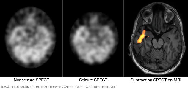
A brain scan can help spot problems in your brain that can sometimes cause epilepsy such as. Common diagnostic tests include electroencephalography EEG which measures electrical activity in the brain and brain scans such as magnetic resonance imaging MRI or computed tomography CT. In patients with epilepsy decreased brain function is seen in the region where seizures originate when the patient is not actually having a seizure.
Which is pretty important when trying to identify epilepsy or indeed many other brain issues. An EEG test gives information about the electrical activity that is happening in your brain at the time the test is carried out. In the United States an estimated 34 million people have epilepsy.
Forty-four patients with epilepsy and 16 healthy control subjects underwent an electroencephalography-correlated functional magnetic resonance imaging study during an eyes-closed condition. Brain imaging is fundamental to the evaluation of people with epilepsy to identify any underlying structural brain abnormality that may be the cause of the epilepsy and to identify a treatment need such as for a tumour vascular disease or infection. However these scans are incredible and play a key part in helping diagnose and treat epilepsy.
An unusual growth brain tumour damage to the brain such as damage caused by a. An MRI scan looks at the structure of the brain and may help to find the cause of. T1 tse coronal oblique 2mm epilepsy protocol Plan the coronal high resolution slices on the sagittal plane.
Angle the position block plane perpendicular to the long axis of the hippocampus. Check the positioning block in the other two planes. Many people with epilepsy only have unusual electrical activity in their brain when they are having a seizure.
Epilepsy is a brain condition that causes patients to fit and lose consciousness. Some of the scans can be noisy MRI fMRI the EEG needs your scalp to be scratched a bit sounds strange right and some need you to go into a little tunnel for a period of time all scans bar the EEG and MEG. Brain imaging has a crucial role in the presurgical assessment of patients with epilepsy.
On the other hand if the patient has a seizure during the test increased brain function is seen.
 Temporal Lobe Epilepsy Epilepsy Foundation
Https Www Ilae Org Files Ilaeguideline Recommendationsforuseof Structuralmri Bernasconi Et Al 2019 Epilepsia Pdf
 Novel Brain Mri Techniques To Improve Epilepsy Surgery Updates From Aes 2018 Consult Qd
 Brain Imaging In Epilepsy Practical Neurology
 Children S Brains Reorganize After Epilepsy Surgery To Retain Visual Perception National Institutes Of Health Nih
 Epilepsy Diagnosis And Treatment Mayo Clinic
Brain Imaging In Epilepsy Practical Neurology
 The Radiology Assistant Role Of Mri
 36 Year Old Woman With Epilepsy From 15 Years Two Axial Mri Scans In Download Scientific Diagram
 Spect Epilepsy Foundation
 Mri And Eeg Could Identify Children At Risk For Epilepsy After Febrile Seizures National Institutes Of Health Nih
 A New Frontier For Epilepsy Brain Imaging Florey
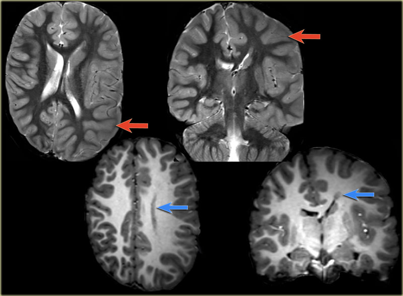
 Characteristics Of Seizure Induced Signal Changes On Mri In Patients With First Seizures Seizure European Journal Of Epilepsy
 Imaging In Epilepsy Journal Of Neurology Neurosurgery Psychiatry


No comments:
Post a Comment
Note: Only a member of this blog may post a comment.