This is to help show up the tissues close to the area containing cancer for example blood vessels. These images are more detailed than regular X-rays.
 Fig 4 4 Chest Radiograph A And Axial Diseases Of The Chest Breast Heart And Vessels 2019 2022 Ncbi Bookshelf
They can give more details about injuries or diseases of.
Chest ct scan. A computed tomography CT scan is a type of imaging test. A CT scan of the chest can discover masses tumors infections or injuries. They can give more information about injuries or diseases of the chest organs.
Like traditional x-rays it produces multiple images or pictures of the inside of the body. Chest CT scans are an important diagnostic tool for diseases such as COVID-19 Some doctors recommend mandatory CT scans of COVID patients to help them with early and better treatment options New Delhi. A lung CT scan short for computed tomography is a type of radiographic scan that uses x-ray technology to create internal images of the chest.
It also can be used to see if cancer has spread into the chest from another area of the body. A CT scan of the chest can help find problems such as infection lung cancer blocked blood flow in the lung pulmonary embolism and other lung problems. A low-dose CT scan is a different type of chest CT scan.
Left lower lobe superior segment. CT Scan of the Chest What is a CT scan of the chest. The technologists will be able to see and hear you at all times during the exam.
A CT examination with an effective dose of 10 millisieverts abbreviated mSv. Commonly CT imaging is used for positioning. Left lower lobe lateral segment.
The radiofrequency waves passing increase the temperature within the met that results in destruction of the tumor. A chest CT scan can help doctors diagnose the cause of shortness of breath or chest pain as well as find the cause of abnormal findings from a standard X-ray. CT scans of the chest You might have an injection of the contrast medium during the scan.
A chest computed tomography CT scan is an imaging test that takes detailed pictures of the lungs and the inside of the chest. Computed tomography more commonly known as a CT or CAT scan is a diagnostic medical imaging test. The scan involves a specialized machine that takes multiple images of your lungs known as slices.
Right lower lobe posterior segment. Right lower lobe anterior segment. A CT scan makes a number of detailed slices pictures which then are merged into one projection.
Computed tomography is an imaging procedure that uses special x-ray equipment to create detailed pictures or scans of areas inside the body. Each picture created during a CT procedure. During a CT scan you lie in a tunnel-like machine while the inside of the machine rotates and takes a series of X-rays from different angles.
The non-contrast CT chest is a commonly performed diagnostic examinationIt is often performed to evaluate conditions impacting the lungs or preferenced over a contrast enhanced scan when iodinated contrast is contraindicated. It is sometimes called computerized tomography or computerized axial tomography CAT. Once the probe is placed in the tumor the radiofrequency energy is delivered.
CT chest is the most important imaging modality in detecting lung changes in COVID-19 infection. If contrast is required the technologist will start an IV in your arm and contrast will be injected through the IV. The CT scan should take only a few minutes.
Chest CT Scan is a more detailed type of chest X-ray that compounds the power of X-rays and computers to deliver a 3D view of your chest. Left lower lobe posterior segment. Learn more about how the test is done and what it can show.
It may help to show whether cancer can be removed with surgery or not. 1 mSv 1 mGy in the case of x-rays may be associated with an increase in the possibility of fatal cancer of. It uses X-rays and a computer to make detailed pictures of the inside of your chest.
These images are better than regular X-rays. It uses X-ray and computer technology to make detailed pictures of the organs and structures inside your chest. A Chest CT scan may help diagnose find.
What Is A Chest CT. The novel coronavirus also known as SARS-CoV-2 causes the COVID-19 disease the infection that has caused the current pandemic. The medical images can help determine if the patient has any of the following disorders3.
CT scans of the chest can identify the cause of various chest symptoms including chest pain cough shortness of breath or fever. The next two pictures show a needle-like RFA probe that is placed through the chest inside the tumor. CT scan is a type of imaging test.
The term tomography comes from the Greek words tomos a cut a slice or a section and graphein to write or record. The cross-sectional images generated during a CT scan can be reformatted in multiple planes. It can be used for Medical Triage.
Computers combine the pictures to create a 3-D model showing the size shape and position of the lungs and structures in the chest. Right lower lobe lateral segment. In a CT scan an X-ray beam moves in a circle around your body.
The CT of the chest requires you to lie down on the scanning table. What is a CT scan of the chest. What is CT Scanning of the Chest.
This creates a clear view that shows the position shape and size of the organs in your chest.
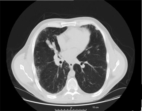
 A 42 Year Old Woman With Abnormal Chest Ct Scan And Chylous Ascites Chest
 Getting Ready For Your Ct Scan Of The Chest Sansum Clinic

 Ct Outperforms Lab Diagnosis For Coronavirus Infection
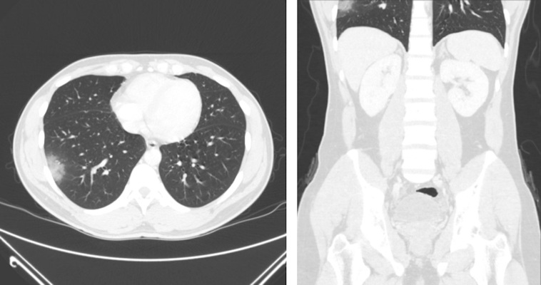 Covid 19 Can Be Seen On Ct Scans Outside The Chest
 Ct Chest Cedars Sinai
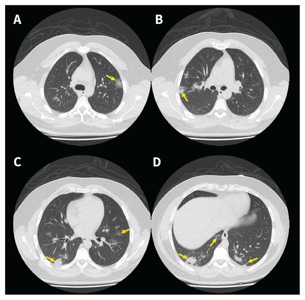
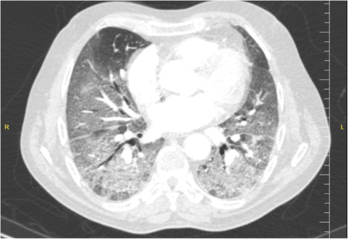
 Ct Manifestations Of Coronavirus Disease 2019 A Retrospective Analysis Of 73 Cases By Disease Severity European Journal Of Radiology
 Ct Scan Database Of 1000 Sets Was Created For Teaching Ai To Diagnose Covid 19 Eurekalert Science News
 Ct Provides Best Diagnosis For Novel Coronavirus Covid 19 Imaging Technology News
 Chest Ct Scan St Elizabeth S Medical Center Steward Family Hospital Brighton Ma
 Chest Ct In Covid 19 What The Radiologist Needs To Know Radiographics
 Clinical And Chest Ct Presentations From 27 Patients With Covid 19 Pneumonia In Mogadishu Somalia A Descriptive Study Egyptian Journal Of Radiology And Nuclear Medicine Full Text
Chest Ct Imaging Of An Early Canadian Case Of Covid 19 In A 28 Year Old Man Cmaj
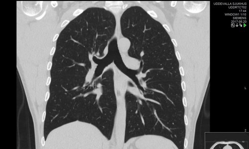
 Normal Ct Chest Radiology Case Radiopaedia Org


No comments:
Post a Comment
Note: Only a member of this blog may post a comment.