Premature Ventricular Complex Trigeminy. Second Degree Heart Block Type I.
 Normal Ecg Beat Types In Record No 100 117 121 212 219 And 234 Download Scientific Diagram
Premature Ventricular Complex Bigeminy.
Types of ekg. An abnormal EKG result can be a sign that one region or section of the heart is. Each ECG lead is presented as a diagram sometimes called a curve. A pre-operative screening ECG is required prior to any surgical procedure that involves general anesthesia because heart disease increases the risk of adverse events from general anesthesia and because this helps your anesthesiologists as they plan your anesthetic medications and surgical.
Adenosine Amiodarone BLS Cardiac arrest Chest pain CPR ECG artifact electrocardiogram Naloxone Resuscitation ST-Segment Elevation STEMI Supraventricular Tachycardia Synchronized Cardioversion takotsubo cardiomyopathy tracheal intubation Wide Complex Tachycardia. Pacemaker Single Chamber Atrial. 1st degree AV block.
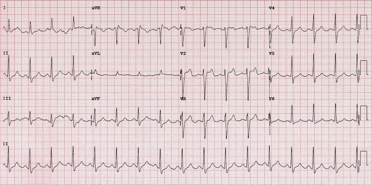
Atrioventricular blocks are a type of heart block that occurs when that impulse is impaired blocked in some way. A standard 12-lead EKG or 12-lead ECG provides information from 12 different areas of the heart as the electrical impulse travels through it. An EKG gives doctors an idea of how hard the heart is working in each specific area.
An ECG is also required prior to any type of heart surgery including surgery for pacemaker placement. Count the number of QRS complexes. Count the number of electrical impulses as represented by PQRST complexes conducted through the myocardium in 60 seconds 1 minute Atrial rate.
Numerous textbooks are devoted to the subject. February 13 2021 Patients often engage in a low impact workout during an EKG test. The main difference between the types is.
For 20 minutes multiple EKG tracings are recorded to evaluate hundreds of cardiac cycles to help detect abnormalities and an increased risk of cardiac arrhythmias. All ECG machines work in basically the same way and are used to perform electrocardiogram tests looking for heart rate abnormalities. An electrocardiogram often abbreviated as ECG or EKG is used to monitor the electrical activity of the heartThere are many different patterns that can be seen in the EKG rhythms that can give vital information to doctors and paramedics.
The types of EKG leads usually correspond to their spatial direction in the results specifically right and left. This version is a more-detailed type of EKG. And anterior and posterior.
The electrocardiograph ECG machine compares amplifies and filters the electrical potential differences recorded by the electrodes and presents the results as ECG leads. The EKG interpretation video series follows along with our EKG interpretation flashcards which are intended to help RN and PN nursing students study for nursing school exams including the ATI HESI and NCLEX. Premature Ventricular Complex Quadrigeminy.
Learn about the different types of electrocardiograms ECG or EKG including holter monitor exercise EKG and resting 12-lead EKG. Typically when you undergo an EKG small electrode. The electrocardiogram ECG or EKG is a diagnostic tool that is routinely used to assess the electrical and muscular functions of the heart.
Supraventricular tachycardia occurs in the upper chambers of your heart known as the atria. While it is a relatively simple test to perform the interpretation of the ECG tracing requires significant amounts of training. It indicates that the atria are contracting pumping blood into the ventricles.
Pacemaker Failure to Pace. Ventricular tachycardia occurs in. These waveforms are labeled P Q R S T and U.
Interpreting EKG Rhythm Strips Step 1 Heart Rate. To briefly summarize the features used in reading EKGs they consist of waveform components which indicate electrical events during one heart beat. P wave is the first short upward movement of the EKG tracing.
There are three subtypes of tachycardia. Electrocardiography ECG or EKG machines are available in a variety of different types that offer different features from the most basic hand-held devices to fully featured machines for use in cardiac centers. Actually analyzing and using the results tends to be a complex science but simply understanding.
Count the number of P waves Ventricular rate.
 Brugada Syndrome Litfl Ecg Library Diagnosis
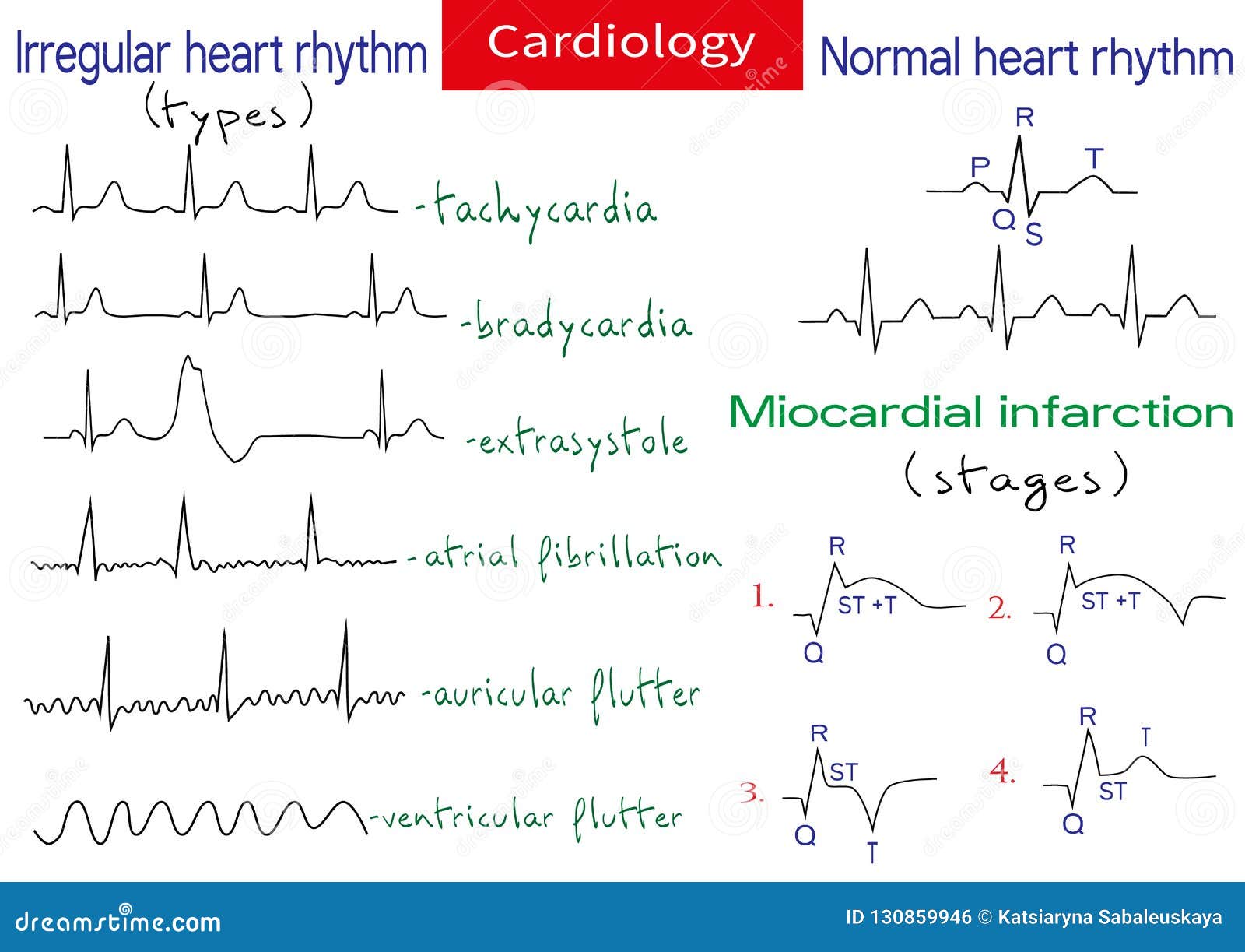
Normal And Pathological Ecg Collection Stock Illustration Illustration Of Cardiac Elevation 130859946
 Different Types Of Ecg Signals Normal Top Arrhythmia Middle And Download Scientific Diagram
 Different Types Of Avnrt Ecg Presentation In All Types The P Wave Is Relatively Narrow In Inferior Leads And In V1 In T P Wave Surface Different Types
 Different Types Of Rhythms Page 1 Line 17qq Com
 The Normal Ecg And Different Types Of Ventricular Arrhythmias A Download Scientific Diagram
 Ecg Interpretation In Brugada Syndrome Sciencedirect
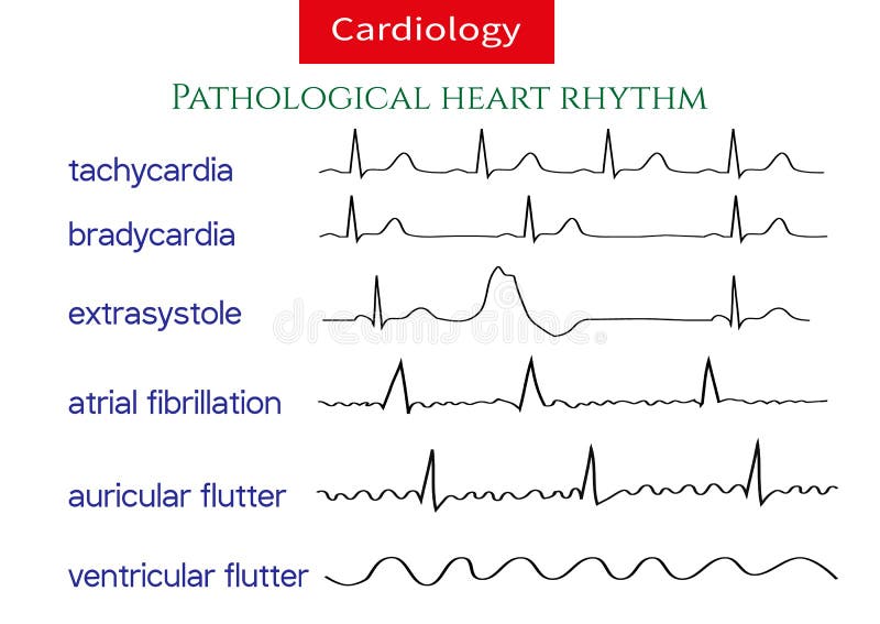 Pathological Ecg Collection Stock Illustration Illustration Of Cardiology Cardiac 130859734
 Pathological Ecg Collection Shematic Vector Illustration Of Different Types Of Irregular Heart Rhythm Shematic Vector Collect Pathology Heart Rhythms Rhythms
 Ecg Interpretation In Brugada Syndrome Sciencedirect
 Illustration Of 16 Types Of Electrocardiogram Ecg Beats Download Scientific Diagram
 Pathological Ecg Vector Photo Free Trial Bigstock
 10 Types Of Ecg Devices For Heart Rhythm Monitoring Medicwiz
 Heart Rhythm Types Page 1 Line 17qq Com


No comments:
Post a Comment
Note: Only a member of this blog may post a comment.