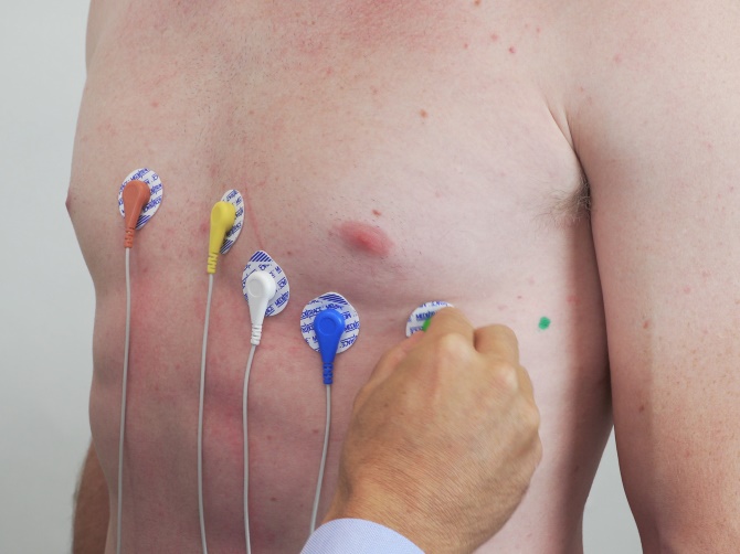
15 LEAD ECG PLACEMENT. Women with larger breasts tissue can displace.
 12 Lead Ecg Placement Guide With Illustrations
The patients chest and all four limbs should be exposed in order to apply the ECG electrodes correctly.

12 lead ecg placement on a female. In both systems the heart is considered an electric dipole. EKG ECG Placement Female - YouTube. The limb leads can also be placed on the upper arms and thighs.
Pocket Reference for the 12-Lead ECG in Acute. An electrocardiogram commonly known as an ECG is a reading that assesses the size and direction of the hearts electrical currents. The Art Of Interpretation Garcia Introduction to 12-Lead ECG PDF.
You can use the technique above if necessary. Similarly an ECG machine also measures repolarization and depolarization of the cardiac muscle cells. The 12-lead ECG is a vital tool for EMTs and paramedics in both the prehospital and hospital setting.
For female patients place leads V3-V6 under the left breast. Additional notes on 12-lead ECG Placement. 12 lead ECG EKG placement of electrode stickers.
In this video I demonstrate electrode placement for recording 12 lead ECGs. There will be a chart on the ECG packaging that lets you know which color corresponds with which lead. EPUB DOWNLOAD 12-Lead ECG.
Explain to the patient what you plan to do in terms of electrode placement. Unpack the ECG leads and read the color-coding system. The 12-lead ECG is used to trace the heart muscle from 12 different electrical positions.
For female patients place leads V3-V6 under the left breast. Electrode Placement V1 - 4th Intercostal space to the right of the sternum V2 - 4th Intercostal space to the left of the sternum V3 - Midway between V2. Despite the appearance of the abdomen during advanced pregnancy placement of the electrodes is the same.
The 15 lead ECG placement is same as 12 lead ECG placement but V4 V5 V6 are placed below the left scapula of the patient on the posterior side. So that an experienced interpreter will see the heart from different angles and make accurate diagnosis. The limb leads can also be placed on the upper arms and thighs.
However there should be uniformity in your placement. The present clinical ECG lead systems the 12-lead system and the VCG systems are results of a long development process of the ECG-theory. While going through nursing school most text diagrams and mannequins show male anatomy.
For instance do not attach an electrode on the right wrist and one on the left upper arm. The leads used in an ECG exam are color coded. This tutorial will demonstrate the lead placement for the 12 lead ECG of the limb leads RA.
The Art of Interpretation Garcia Introduction to 12-Lead ECG FREE PDF. 12-Lead ECG ELECTRODE PLACEMENT FOR PREGNANT PATIENTS. The 12 Lead placement is one of the most productive investigations in medicine.
In case of female patients place V3 and V6 leads under the left breast. 12-Lead ECG Placement. 2021 ConectMed Technologies Co Ltd 2021 Shenzhen Yong Qiang Fu Industry CoLtd.
Note that left-axisdeviation on the ECG may appear in both pregnant. Every electrode will seize the electrical activity from a different position. For instance do not attach an electrode on the right wrist and one on the left upper arm.
However there should be uniformity in your placement. These V4 V5 V6 when placed below the scapula are called as V13 V14 V15. The 6 chest leads are medically referred to as the V leads.
This includes cardiac aulsculation respiration aulsculation locations as well as 4-lead and 12-lead ecg placement. I have a growing playlist of detailed tutorials for reading and interpreting 12. Incorrect placement can lead to a false diagnosis of infarction or negative changes on the ECG.
Additional notes on 12-lead ECG Placement. It is extremely important to know the exact placement of each electrode on the patient. Read The 12-Lead ECG in Acute Coronary Syndromes.
Emphasize that several of the chest leads may need to be placed around and under the left breast. There are different methods for identifying the correct landmarks for ECG electrode placement the two most common being the Angle of Louis Method and the Clavicular Method Crawford Doherty 2010a. At a minimum lead V4 should be placed on the 5th intercostal mid-clavicular exact opposite of the regular left side placement if an inferior infarct was originally seen in leads II III and AVF.
No2 BuildingXinwei Villagethe second industrial zoneDalang StreetLonghua DistrictShenzhenChina. Lastly a right sided 12-lead ECG placement allows you to detect a right sided infarct.


